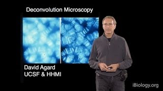
Media Summary: This video demonstrates how to setup, in ImageJ, the Podcast based on Qiao, et al. 2024 ( describing in detail the concepts and ... raw image acquired on a Zeiss imager Z1, using a 63x/1.4NA oil objective, excitation 405nm (cyan) and 640 nm (red), apotome ...

Zero Shot Deconvolution For Microscopy - Detailed Analysis & Overview

This video demonstrates how to setup, in ImageJ, the Podcast based on Qiao, et al. 2024 ( describing in detail the concepts and ... raw image acquired on a Zeiss imager Z1, using a 63x/1.4NA oil objective, excitation 405nm (cyan) and 640 nm (red), apotome ... In this video, learn how to use the FLUOVIEW FV3000 to perform spectral An introduction to principles and practice of Video abstract of the paper A Joint Richardson-Lucy

abberior homepage: abberior shop: abberior TRUESHARP: ... This week features "Overcoming physical resolution limits of fluorescence This video was recorded by the Live Cell Imaging facility at the Karolinska Institute in Sweden during the LCI course 2026. Mouse fibroblast spheroid (NIH 3T3, 500 cells) was imaged in a INCell Analyzer 2500HS ( ). Magnification:4X.

Photo Gallery

Zero-Shot Deconvolution for Microscopy Images
Zero-Shot Deconvolution to Enhance Super-Res Microscopy
Microscopy: Deconvolution Microscopy (David Agard)
Apotome vs Deconvolution for widefield fluorescence
True Deconvolution Webinar (March 10, 2020 | 17:00 CET - 11:00 EST - 08:00 PST)
FV31S-SW Image Processing: Spectral Deconvolution
Deconvolution deconvolved
Video abstract Joint Richardson-Lucy Deconvolution Algorithm for mSIM
TRUESHARP image boosting – deconvolution you can trust! Part 2
Liangyi Chen - Overcoming physical resolution limits with sparse deconvolution - Imaging ONEWORLD
What does deconvolution microscopy mean?
Introduction to 2D and 3D deconvolution   2026
View Detailed Profile
Zero-Shot Deconvolution for Microscopy Images

Zero-Shot Deconvolution for Microscopy Images

This video demonstrates how to setup, in ImageJ, the

Zero-Shot Deconvolution to Enhance Super-Res Microscopy

Zero-Shot Deconvolution to Enhance Super-Res Microscopy

Podcast based on Qiao, et al. 2024 (https://www.nature.com/articles/s41467-024-48575-9) describing in detail the concepts and ...

Microscopy: Deconvolution Microscopy (David Agard)

Microscopy: Deconvolution Microscopy (David Agard)

https://www.ibiology.org/talks/

Apotome vs Deconvolution for widefield fluorescence

Apotome vs Deconvolution for widefield fluorescence

raw image acquired on a Zeiss imager Z1, using a 63x/1.4NA oil objective, excitation 405nm (cyan) and 640 nm (red), apotome ...

True Deconvolution Webinar (March 10, 2020 | 17:00 CET - 11:00 EST - 08:00 PST)

True Deconvolution Webinar (March 10, 2020 | 17:00 CET - 11:00 EST - 08:00 PST)

The essence of using

FV31S-SW Image Processing: Spectral Deconvolution

FV31S-SW Image Processing: Spectral Deconvolution

In this video, learn how to use the FLUOVIEW FV3000 to perform spectral

Deconvolution deconvolved

Deconvolution deconvolved

An introduction to principles and practice of

Video abstract Joint Richardson-Lucy Deconvolution Algorithm for mSIM

Video abstract Joint Richardson-Lucy Deconvolution Algorithm for mSIM

Video abstract of the paper A Joint Richardson-Lucy

TRUESHARP image boosting – deconvolution you can trust! Part 2

TRUESHARP image boosting – deconvolution you can trust! Part 2

abberior homepage: https://abberior.rocks abberior shop: https://abberior.shop abberior TRUESHARP: ...

Liangyi Chen - Overcoming physical resolution limits with sparse deconvolution - Imaging ONEWORLD

Liangyi Chen - Overcoming physical resolution limits with sparse deconvolution - Imaging ONEWORLD

This week features "Overcoming physical resolution limits of fluorescence

What does deconvolution microscopy mean?

What does deconvolution microscopy mean?

What does

Introduction to 2D and 3D deconvolution   2026

Introduction to 2D and 3D deconvolution 2026

This video was recorded by the Live Cell Imaging facility at the Karolinska Institute in Sweden during the LCI course 2026.

Deconvolution of images of cell spheroid imaged by fluorescence microscopy

Deconvolution of images of cell spheroid imaged by fluorescence microscopy

Mouse fibroblast spheroid (NIH 3T3, 500 cells) was imaged in a INCell Analyzer 2500HS (@ipencnen ). Magnification:4X.