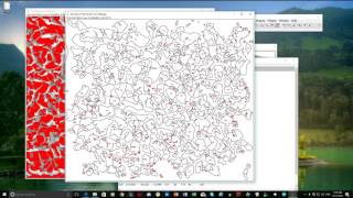
Media Summary: Digital analysis coral fragments Now that you have taken the pictures of the coral fragments, you can analyse them with All you need to know for getting started with This is the second part of the explanation on how to use
Tutorial Imagej V2 - Detailed Analysis & Overview
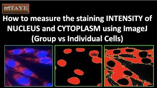
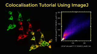
Digital analysis coral fragments Now that you have taken the pictures of the coral fragments, you can analyse them with All you need to know for getting started with This is the second part of the explanation on how to use How to measure the staining INTENSITY of NUCLEUS and CYTOPLASM using ImageJ Software Learn how to use 3D visualization & animation tools offered by FIJI ( Fiji is the most important image analysis tool for the biologist! In this video, learn how to open and import images from microscopes ...
Follow us on Instagram: ⭕ Subscribe: It takes a lot of time in creating these videos, ... This video was made by Maryville College Biology student Lauren Evans as part of her Senior Study. It shows the procedure for ...