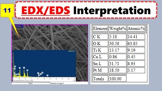
Media Summary: We've talked a lot about light microscopy, but this technique has inherent limitations in resolution and magnification. The next ... These are also referred to as point scans and so because we have a scanning electron beam in the Examining a metal coil and surface oxidation using the Phenom Pro X

Sem Micrographs Interpretation In Experimental - Detailed Analysis & Overview

We've talked a lot about light microscopy, but this technique has inherent limitations in resolution and magnification. The next ... These are also referred to as point scans and so because we have a scanning electron beam in the Examining a metal coil and surface oxidation using the Phenom Pro X Connector terminals of a worn SD card were observed by Learn how magnification and resolution differ in

Photo Gallery

SEM Micrographs Interpretation in Experimental paper:  Scanning Electron Microscopy SEM Analysis
Electron Microscopy (TEM and SEM)
How to Read SEM Images | Microstructure Analysis Explained
MSE585 F20 Lecture 16 Module 5 - SEM-EDS Scanning Modes
SEM Cross Section Analysis | How to Interpret Micrographs
SEM/EDS: Inserting Samples into the SEM
How to measure the surface roughness from SEM image using Image J software
Metal Oxidation - Part 1: SEM Imaging of a Metal Coil (NVCC 9/13)
HOW TO Focus A Noisy SEM Image || Pro SEM Tip!
[Electronics] SEM analysis of an SD card connector terminal contamination and scratches
EDS/EDX Microsctructure Interpretation: Energy -Dispersive X-rays Spectroscopy Analysis
SEM Images: Magnification vs Resolution (Clear Difference Explained)
View Detailed Profile
SEM Micrographs Interpretation in Experimental paper:  Scanning Electron Microscopy SEM Analysis

SEM Micrographs Interpretation in Experimental paper: Scanning Electron Microscopy SEM Analysis

How to interpret

Electron Microscopy (TEM and SEM)

Electron Microscopy (TEM and SEM)

We've talked a lot about light microscopy, but this technique has inherent limitations in resolution and magnification. The next ...

How to Read SEM Images | Microstructure Analysis Explained

How to Read SEM Images | Microstructure Analysis Explained

Learn how to interpret

MSE585 F20 Lecture 16 Module 5 - SEM-EDS Scanning Modes

MSE585 F20 Lecture 16 Module 5 - SEM-EDS Scanning Modes

These are also referred to as point scans and so because we have a scanning electron beam in the

SEM Cross Section Analysis | How to Interpret Micrographs

SEM Cross Section Analysis | How to Interpret Micrographs

Learn how to interpret

SEM/EDS: Inserting Samples into the SEM

SEM/EDS: Inserting Samples into the SEM

To insert your sample in the

How to measure the surface roughness from SEM image using Image J software

How to measure the surface roughness from SEM image using Image J software

How to measure the surface roughness of

Metal Oxidation - Part 1: SEM Imaging of a Metal Coil (NVCC 9/13)

Metal Oxidation - Part 1: SEM Imaging of a Metal Coil (NVCC 9/13)

Examining a metal coil and surface oxidation using the Phenom Pro X

HOW TO Focus A Noisy SEM Image || Pro SEM Tip!

HOW TO Focus A Noisy SEM Image || Pro SEM Tip!

Contact me for

[Electronics] SEM analysis of an SD card connector terminal contamination and scratches

[Electronics] SEM analysis of an SD card connector terminal contamination and scratches

Connector terminals of a worn SD card were observed by

EDS/EDX Microsctructure Interpretation: Energy -Dispersive X-rays Spectroscopy Analysis

EDS/EDX Microsctructure Interpretation: Energy -Dispersive X-rays Spectroscopy Analysis

How to interpret EDS/EDX

SEM Images: Magnification vs Resolution (Clear Difference Explained)

SEM Images: Magnification vs Resolution (Clear Difference Explained)

Learn how magnification and resolution differ in

SEM/EDS: Making EDS Analyses Part 3

SEM/EDS: Making EDS Analyses Part 3

SEM/EDS: Making EDS Analyses Part 3