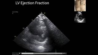
Media Summary: In this 16-minute video, Dr Katie Wiskar explains how to use Point-of-Care Ultrasound to estimate Time to talk LVEF—the heartbeat of cardiac Book a 1-on-1 Clarius demo: E-point septal separation (EPSS) is an easy way to quickly and ...
Pocus For Lv Systolic Function - Detailed Analysis & Overview
In this 16-minute video, Dr Katie Wiskar explains how to use Point-of-Care Ultrasound to estimate Time to talk LVEF—the heartbeat of cardiac Book a 1-on-1 Clarius demo: E-point septal separation (EPSS) is an easy way to quickly and ... Book a 1-on-1 Clarius demo: In this 1-hour webinar, Dr. Cook demonstrates how to use Dr. Golzar shares her knowledge about cardiac ultrasound. Book a 1-on-1 Clarius demo: In this video Dr. Cook performs a quick assessment of
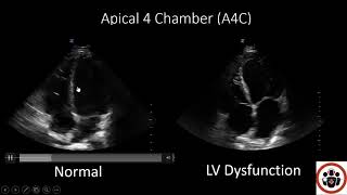
Step by Step guide to using M-mode on Ultrasound. E Point Septal Separation (EPSS) Measurement used as an example. Book a 1-on-1 Clarius demo: Tricuspid annulus plane After watching this video, you will be able to recognize normal, hyperkinetic, and hypokinetic After watching this video, you will be able to recognize right ventricular dilatation, significant hypertrophy, and hyper- and ...